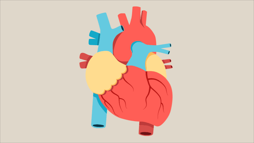
The heart is a muscular organ that lies at the center of the circulatory system. It is the pump that powers your body by delivering blood through a network of chambers, veins, and arteries. About the size of your fist, the heart’s function is essential to human life. Therefore, it is important to know how this vital organ functions. Let’s start with the basic anatomy!
What are the basic parts of the heart?
The heart consists of four chambers- two on top and two on the bottom. The chambers on top are called atriums while the chambers on the bottom are called ventricles. When looking at the anterior view of the heart, the “left” chambers are actually referred to as the right and the “right” chambers are called the left. Ventricle chambers pump blood out of the heart and are separated by a wall known as the interventricular septum. The atria (right and left atrium) receive blood that enters the heart and are separated by a wall known as the interatrial septum.
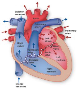
Aside from the chambers, the heart contains four valves. Valves contain leaflets and act as one-way inlets and outlets for blood flowing into and out of the ventricle. Leaflets are sturdy, thin flaps of tissue that open to allow blood flow from one chamber to another during half of the heartbeat and close to prevent blood backflow during the second half of the heartbeat. Located between the right ventricle and right ventricle lies the tricuspid valve. The tricuspid valve contains three leaflets. The pulmonary valve is located between the right ventricle and the pulmonary artery. This mitral valve is located between the left atrium and the left ventricle and contains two leaflets unlike the tricuspid. Lastly, the aortic valve is located between the left ventricle and aorta. The heart is surrounded by a sac-like double layered membrane known as the pericardium. The outer layer is fibrous pericardium It is made of thick connective tissue and is attached by ligaments to the diaphragm, spinal column, and other parts of the body. The inner layer is serous pericardium and is further divided into the viscera and parietal layers. The inner layers of the pericardium are attached to the heart. Between the two layers lies the fluid filled pericardial cavity. The cavity facilitates lubrication and free movement of the heart.
How does blood flow through the heart?
Blood generally flows through the heart in four basic steps. Oxygen poor blood enters the right atrium and is pumped into the right ventricle through the tricuspid valve. The right ventricle then pumps the oxygen poor blood into the lungs via the pulmonary valve.
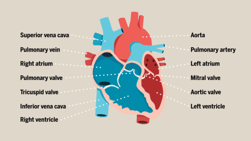
The new oxygen rich blood is received by the left atrium from the lungs. This oxygen rich blood is then pumped through the mitral valve to the left ventricle. The left ventricle then pumps the oxygen rich blood through the aortic blood to the aorta, the main artery which carries blood away from the heart and to the rest of the body.
Check out the next post to learn about the cardiac electrical conduction system!
Resources
Content – Health Encyclopedia – University of Rochester Medical Center, http://www.urmc.rochester.edu/encyclopedia/content.aspx?ContentTypeID=90.
“Default – Stanford Children’s Health.” Stanford Children’s Health – Lucile Packard Children’s Hospital Stanford, http://www.stanfordchildrens.org/en/topic/default?id=anatomy-and-function-of-the-heart-valves-90-P03059.
“Heart Information Center: Heart Anatomy.” Texas Heart Institute, 2 Feb. 2020, http://www.texasheart.org/heart-health/heart-information-center/topics/heart-anatomy/.
“How the Heart Works.” How the Heart Works | Michigan Medicine, http://www.uofmhealth.org/health-library/tx4097abc.
“How the Heart Works.” MyHealth.Alberta.ca Government of Alberta Personal Health Portal, myhealth.alberta.ca/Health/pages/conditions.aspx?hwid=tx4097abc.
Medicine, Michigan. “Anatomy of a Human Heart.” Health & Wellness Topics, Health Tips & Disease Prevention, 27 Feb. 2019, healthblog.uofmhealth.org/heart-health/anatomy-of-a-human-heart.
